
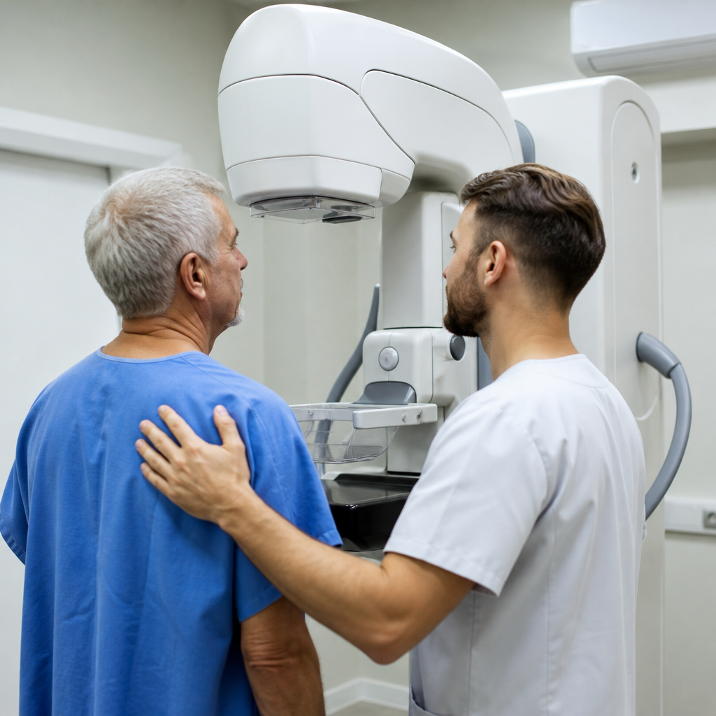
The transition to Medicare represents a significant shift in how healthcare decisions are made, particularly when it comes to diagnostic procedures that require pre-authorization or meet specific medical necessity criteria. For women navigating breast health concerns during this transition, understanding which imaging technologies are available and when they become appropriate clinical tools can prevent delays in diagnosis and ensure comprehensive care planning.
Breast cancer screening remains one of the most critical preventive services covered under Medicare Part B, yet confusion persists about the various imaging modalities available beyond standard mammography. As medical technology advances and detection methods become more sophisticated, patients entering the Medicare system often face choices they did not encounter with their previous insurance coverage.
The Standard Screening Pathway Under Medicare
Medicare Part B covers annual screening mammograms for all women aged 40 and older, with no copayment or deductible when performed by a provider who accepts assignment. This baseline coverage provides the foundation for breast health monitoring, but it represents only one tool in a broader diagnostic arsenal. The screening mammogram serves as the first-line approach for detecting abnormalities in asymptomatic women, typically using digital X-ray technology to create images of breast tissue.
When a screening mammogram reveals an area of concern, or when a woman presents with symptoms such as a palpable lump, nipple discharge, or skin changes, the diagnostic pathway expands. Diagnostic mammograms, which involve additional views and magnification of specific areas, are also covered under Medicare Part B. These targeted examinations allow radiologists to characterize findings more thoroughly before determining whether additional imaging is warranted.
When Advanced Imaging Becomes Medically Necessary
Certain clinical scenarios call for imaging techniques that provide different types of information than conventional mammography can deliver. Women with extremely dense breast tissue, for instance, may have reduced mammographic sensitivity because dense fibroglandular tissue can obscure small lesions on X-ray images. Similarly, patients with genetic predispositions to breast cancer, such as BRCA1 or BRCA2 mutations, may benefit from supplemental screening approaches that offer enhanced detection capabilities.
Magnetic resonance imaging has emerged as a valuable complement to mammography in specific high-risk populations. Unlike mammography, which uses ionizing radiation to create images, this technology employs powerful magnetic fields and radio waves to generate detailed cross-sectional images of breast tissue. For patients seeking advanced diagnostic evaluation, facilities such as Cleveland Clinic Abu Dhabi offer comprehensive mri for breast imaging services that can detect abnormalities not visible on conventional mammograms, particularly in women with dense breast tissue or elevated genetic risk profiles.
Medicare coverage for advanced breast imaging typically requires documentation of medical necessity. This means a physician must establish that standard mammography is insufficient for adequate evaluation given the patient’s individual risk factors, breast density, or suspicious findings that require further characterization. The authorization process ensures that these more expensive and time-intensive procedures are directed toward patients who will derive the greatest clinical benefit.
Navigating Medicare Advantage Plans and Imaging Coverage
Many Medicare beneficiaries choose Medicare Advantage plans as an alternative to Original Medicare, and these private insurance options often handle diagnostic imaging coverage differently. Understanding what to expect from Medicare Advantage can help patients anticipate potential requirements such as prior authorization for advanced imaging studies, network restrictions that limit where procedures can be performed, and referral requirements that may differ from Original Medicare rules.
Medicare Advantage plans must cover at minimum everything Original Medicare covers, but they may impose additional utilization management protocols. For breast imaging, this often means that while screening mammograms remain straightforward to obtain, diagnostic mammograms and advanced imaging modalities may require pre-authorization from the plan’s medical review team. Patients should verify their specific plan’s requirements before scheduling any diagnostic procedure beyond routine screening.
The Role of Academic Medical Centers in Imaging Quality
Research consistently demonstrates that the quality of diagnostic imaging interpretation improves in settings where radiologists regularly encounter complex cases and participate in ongoing education. Academic and teaching hospitals typically maintain higher volumes of advanced imaging studies, which allows their radiology departments to develop expertise in detecting subtle abnormalities and distinguishing benign findings from those requiring biopsy or further investigation.
Studies have shown that the presence of teaching hospitals improves outcomes across multiple clinical domains, including diagnostic accuracy in radiology. For Medicare beneficiaries facing breast imaging decisions, seeking care at facilities with academic affiliations or those that participate in radiologist training programs may offer advantages in terms of subspecialty expertise and access to the latest imaging protocols.
Preparing for Diagnostic Imaging Appointments
Once advanced breast imaging has been scheduled, patients can take several steps to ensure the procedure yields the most useful diagnostic information. Bringing previous mammogram images and reports allows the radiologist to compare current findings with historical baselines, which can be crucial for determining whether a finding is new or has been stable over time. Many imaging centers can request these records directly from previous providers, but patients should confirm this arrangement at least one week before their appointment.
For procedures involving contrast agents, such as magnetic resonance imaging with gadolinium enhancement, patients should inform their care team about kidney function, as impaired renal clearance can affect contrast safety. Medicare typically covers the lab work necessary to assess kidney function before contrast-enhanced studies, but this testing must be ordered by the referring physician and completed in advance of the imaging appointment.
Cost Considerations and Medicare Part B Deductibles
While screening mammograms are fully covered with no cost-sharing under Medicare Part B, diagnostic imaging studies are subject to the annual Part B deductible and the standard 20 percent coinsurance. For 2024, the Part B deductible is $240, after which Medicare pays 80 percent of the Medicare-approved amount for covered services. This means that a diagnostic mammogram or advanced imaging study could result in out-of-pocket costs ranging from several dozen to several hundred dollars, depending on when in the calendar year the service is performed and whether the deductible has already been met through other Part B services.
Patients concerned about these costs should discuss payment options with the imaging facility’s billing department before the procedure. Some facilities offer payment plans or financial assistance programs for Medicare beneficiaries with limited income. Additionally, Medigap supplemental insurance policies can cover most or all of the Part B coinsurance and deductible, substantially reducing out-of-pocket expenses for diagnostic procedures.
Following Through on Imaging Results
The diagnostic imaging process does not end when the procedure is complete. Radiologists typically issue preliminary readings within 24 to 48 hours, with final reports following shortly thereafter. These reports are sent to the ordering physician, who is responsible for communicating results to the patient and recommending next steps. Medicare beneficiaries should establish clear communication channels with their primary care provider or specialist to ensure they receive results promptly and understand what follow-up is needed.
When imaging reveals findings requiring tissue sampling, Medicare Part B covers breast biopsies as medically necessary diagnostic procedures. The type of biopsy performed depends on the size, location, and characteristics of the abnormality, with options ranging from ultrasound-guided core needle biopsy to stereotactic or magnetic resonance-guided procedures for lesions visible only on specific imaging modalities. Understanding that Medicare coverage extends through the entire diagnostic cascade can provide reassurance during what is often an anxious waiting period.
Breast health management under Medicare requires active patient participation in understanding coverage rules, advocating for appropriate imaging when clinical circumstances warrant it, and ensuring continuity across the diagnostic process. By familiarizing themselves with the available imaging technologies and the criteria that make advanced studies medically necessary, Medicare beneficiaries can work effectively with their healthcare teams to obtain thorough evaluation while navigating the insurance landscape efficiently.